Incredible Inner Space Exhbition by Microscopy Australia
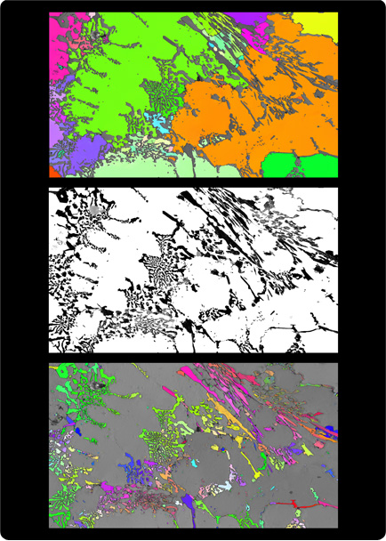 Crystals in an innovative super-hard cast iron. White and black show regions of different chemical composition within the iron. The adjacent images show the orientation of the crystals in the white region (top) and the black region (bottom).
Visualised using electron backscatter diffraction by Chris Barry, Nick Ferguson, Dr Pat Trimby, Dr Gwénaëlle Proust and A/Prof. Julie Cairney, University of Sydney.
Size: 253 micrometres wide.
This cast iron is super hard – it has to be. It is used in mines to push around crushed-up rocks so the minerals can be extracted. Weir minerals develop their own types of cast iron that are even harder than most. This kind of image helps the researchers to understand more about the structure of the iron and how it changes when it undergoes different processes. The harder the iron, the longer the machines last. It can cost millions of dollars a day to stop and replace the machines so the less often you have to do it, the less it costs the mine.


