Incredible Inner Space Exhbition by Microscopy Australia
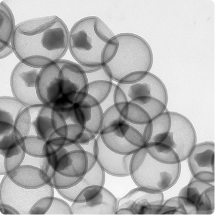 Hollow silica nano-spheres being developed for drug delivery. The drug seeps into the spheres through pores in the silica, which will then be blocked until they are ready to be opened inside the patient's body.
Visualised using transmission electron microscopy by Sarah Renner, University of New South Wales.
Size: each sphere is 125 nanometres across.
Transmission electron microscopy allows the production of these spheres to be monitored. The spheres are solid to begin with and the cores are gradually dissolved using a chemical called sodium borohydride. Here, most of the hollow spheres still contain smaller core particles, showing that the process is not yet finished.


