Supported by the International Job Shadowing Program, Microscopy Australia expert Dr Chad Moore recently attended the EMBO-sponsored Deep Learning for Microscopy Image Analysis course in Italy to expand his knowledge of AI-assisted image analysis. The course covered a wide range of deep learning approaches for dealing with diverse microscopy image datasets including deep learning models, image regression and restoration, segmentation and more, all using open-source software.
Dr Moore is now sharing these learnings with the wider Australian research community. He recently presented a webinar, ‘Deep learning denoising for volume microscopy’ (watch it on YouTube) with one of the instructors from the course, Dr Ben Salmon, to Volume Imaging Australia, a special interest group of the Australian Microscopy and Microanalysis Society. This illustrates not only the value of the learnings, but also of the international connections made during the job shadowing program.
“It was invaluable to be present in person to pick the brains of those developing the open-source solutions that we can all utilise, from getting advice on approaching particular types of data to insights into where the field is heading over the coming 2–3 years” – Dr Chad Moore, The University of Sydney
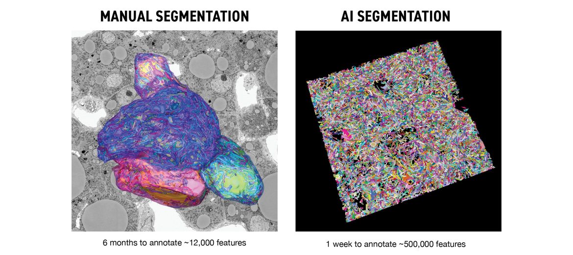
Also part of the Sydney Microscopy and Microanalysis team, Dr Gerry Shami has been working on improving image analysis efficiency in volume electron microscopy (VEM). VEM allows ultra-high resolution 3D imaging of biological materials. It also produces notoriously large datasets that are time-intensive to analyse. Dr Shami’s deep learning approach, developed in collaboration with Zeiss, led to an almost ~1,000-fold increase in analysis speed. Previously, it took six months to annotate approximately 12,000 features. Now, with this new method, they can annotate around 500,000 features in just one week.
“Undoubtedly, AI solutions are having a tremendous impact not only in volume electron microscopy but microscopy more broadly. I believe the greatest advantages are the large volumes that can be probed and the high throughput that can be obtained. These factors ultimately lead to meaningful quantitative results that would otherwise be unachievable with conventional approaches” – Dr Gerry Shami, The University of Sydney
Dr Gerry Shami had ~1000-fold increase in analysis speed using AI tools to complete segmentation, compared to previous manual methods. Data set is of mitochondria in human metabolic fatty liver disease imaged using volume electron microscopy at Microscopy Australia's University of Sydney facility.
December 4, 2024