The Editor said “Scientific Reports published more than 1,940 cell and molecular biology papers in 2021, and so a position in the top 100 most downloaded articles is an extraordinary achievement — your science is of real value to the research community.” Published in February 2021, their paper received 3,897 article downloads in the first nine months.
The work described changes in the structure of mitochondria in cells from patients with non-alcoholic fatty liver disease (NAFLD). Mitochondria are cellular structures that generate the energy required to power the cell’s biochemical reactions. Peculiarly shaped, giant mitochondria are often seen in liver biopsies from patients with NAFLD; those exposed to toxins, anti-cancer drugs, free-radicals, or nutritional deficiencies; or as a consequence of high-fat Western diets and a variety of other diseases.
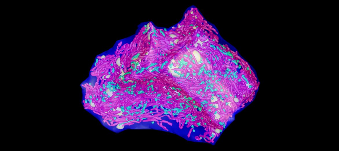
Our expert staff, Prof. Filip Braet and Dr Gerald Shami, along with researchers, used an advanced 3D multimodal imaging approach at Microscopy Australia’s University of Sydney facility to image and measure thousands of mitochondria. They showed that the highly folded membranes inside normal mitochondria were more irregular with far fewer folds in the giant mitochondria. These giants also contained granules, crystalline inclusions and voids, not seen in normal mitochondria.
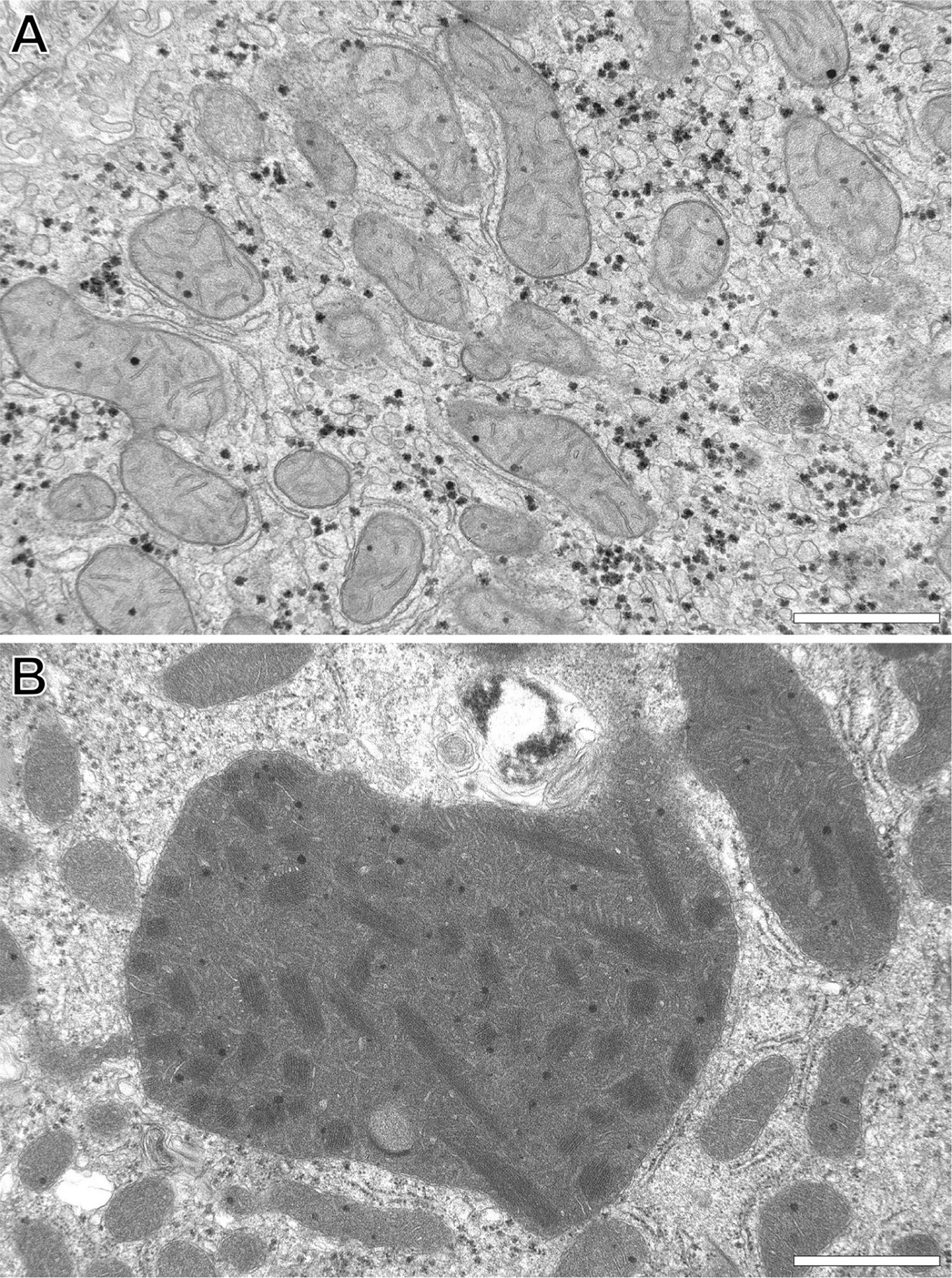
Transmission electron microscopy images of (A) normal human liver mitochondria versus (B) which shows a giant mitochondrion that is many times larger relative to normal mitochondria and displays a distinctly altered internal fine structural appearance. The scale bar = 1 micrometre.
The 3D nature of the techniques used, made it possible to measure mitochondrial surface area and volumes as well as understanding the structures and arrangements of the inclusions. This led the researchers to suggest a model where mitochondria fuse to form the giant mitochondria seen in the disease state. Further advanced techniques available in the University of Sydney facility are now being applied to the analysis of the inclusions seen in giant mitochondria to get a better understanding of what they are made of and how they relate to disease.
G. Shami et al. 2021 Scientific Reports DOI: 10.1038/s41598-021-82884-z
3D image of a liver cell with the normal mitochondria in green and the giant mitochondria in red generated by Transmission electron tomography.
March 25, 2022