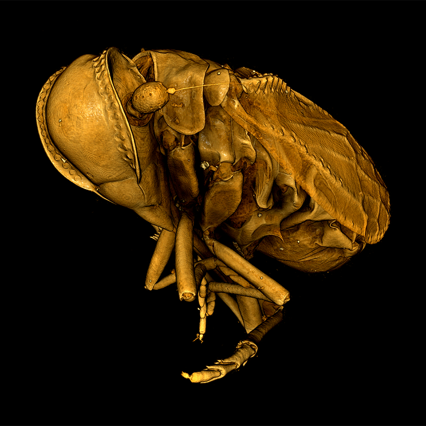
Microscopy Australia’s micro-CT facilities at the Australian National University (ANU) and the University of Western Australia (UWA) are each helping to reveal hidden samples and see inside valuable specimens without damaging them. The Australian Museum in Sydney selects high priority specimens (those in demand for researchers, difficult to assess through photos, challenging to ship, and sometimes the ‘type’ specimens on which species are based) and carefully sends them to the CTLab at ANU. There, each specimen is imaged in micro-CT instruments. The data is reconstructed into a 3D volume with interactive surfaces, destined for the Australian Museum’s online portal that uses Pedestal, a product produced by Macquarie University. The portal’s digital archives will let visitors interact with specimens that would not otherwise be on display.

Micro-CT reconstruction of Phaconeura proserpina, a blind cave dwelling plant-hopper, taken at Microscopy Australia’s University of Western Australia facility.
The Western Australian Museum is also undertaking a similar project, using our Micro-CT facilities at UWA to create full 3D datasets of insects, crustaceans, arachnids and fossil shark teeth that will be turned into interactive digital displays or larger-than-life scale models.
“With micro-CT imaging we can bring the microscopic world to life at a scale that people can appreciate.” – Entomology curator Dr Nikolai Tatarnic, Western Australian Museum
As a result of these scans, visitors can see the microscopic details of the minuscule creatures that make up most of the animal diversity on the planet, inspiring children and adults with the wonder of science and nature. Researchers can visualise previously inaccessible morphological data without destroying precious samples. A more complete set of specimens is available for the public to explore, bringing added benefits to people in regional centres.
Micro-CT reconstruction of Occiperipatoides gilesii, a velvet worm from the southwest of Western Australia, taken at Microscopy Australia's University of Western Australia Facility, the Centre for Microscopy, Characterisation, and Analysis (CMCA).
November 28, 2019