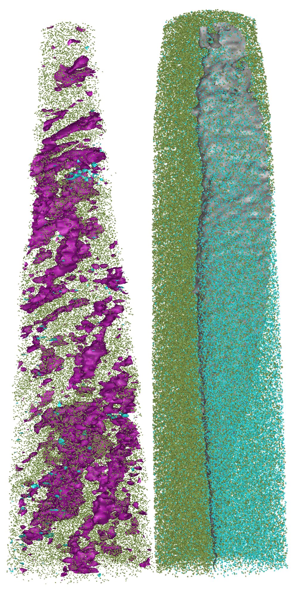
APT reconstructions showing the placement of atoms within normal bone (left) and the implant (right) after being implanted in a sheep for 12 months. Purple is carbon, green is calcium and turquoise is aluminum. At it’s thinnest point, left is~20 nm across, while right is ~30 nm across.
Large bone defects and loss due to cancer or trauma can result in scar tissue that impairs the bones’ ability to repair and regenerate even with current scaffolds. Developing scaffolds that are strong enough for use in weight-bearing bones such as those in the leg or spine is especially challenging.
By using Microscopy Australia’s University of Sydney facilities, Prof. Hala Zreiqat’s research team developed an innovative synthetic bioceramic bone scaffold to overcome these problems. It incorporates trace elements that encourage bone growth and is strong enough for use in load-bearing bones. However, the researchers needed a technique that would allow them to confirm:
Atom probe tomography (APT) has long been used in engineering and material sciences to examine the 3D composition of a material, such as a metal, at the atomic level. However, it has rarely been used on more structurally complex biological samples. Microscopy Australia technique development specialist Dr Natalie Holmes has been working with atom probe manufacturer CAMECA, and Prof. Zreiqat’s team, to develop ways to use APT so it can offer insight into the processes of biodegradation and integration of synthetic bone implants after incorporation into the body. Several other expert staff from Microscopy Australia also contributed to this project including NanoSIMS expert Dr Paul Guagliardo and atom probe expert Prof. Julie Cairney.
The implant was tested in a 12-month animal trial where it was implanted into sheep tibia. After this trial, samples of the bone scaffold, the newly formed tissue, and the surrounding original bone were removed and examined using APT. Biological tissue, even one as hard as bone, is very challenging to image using APT. After a lot of experimenting, optimising instrument settings, testing sample preparation methods and working out how to measure the atoms they needed to detect, the team arrived at a reliable work flow that generated consistent results.
The tomography revealed the incorporation of trace elements from the bioceramic implant into the new tissue structure, demonstrating that it could be supporting new bone growth. APT also revealed that the implant slowly disappeared as the new bone took over.
This new application of APT has addressed a gap in biomedical imaging methods, enabling researchers to examine the composition of bone at the atomic scale. This allows for better implant design, resulting in better outcomes for the cancer and trauma patients who need them. It will also be relevant to other research groups working to better understand bone diseases.
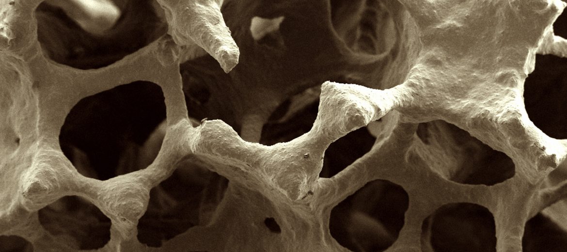
Scanning electron micrograph of bone scaffold imaged at the University of Sydney
Reference
Holmes, N. P., Roohani, I., Entezari, A., Guagliardo, P., Mirkhalaf, M., Lu, Z., Chen, Y.-S., Yang, L., Dunstan, C. R., Zreiqat, H., & Cairney, J. M. (2023). Discovering an unknown territory using atom probe tomography: Elemental exchange at the bioceramic scaffold/bone tissue interface. Acta Biomaterialia, 162, 199–210.
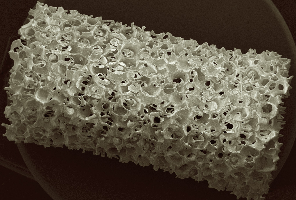
A scanning electron microscope image of the bone implant devleoped by Prof. Hala Zreiqat's research team.
January 23, 2024