RBCs develop in the bone marrow in cell clusters called erythropoietic islands. Prof. Stuart Fraser from the University of Sydney (USyd) has been studying the relationship of island structure to the development of RBCs.
Different proteins are present on the surfaces of RBCs at different stages of their development. These proteins can be labeled to identify the developmental stage of any particular cell. By attaching tiny gold nanospheres in turn to each of the unique molecules it is possible to determine the developmental stage of the RBCs.
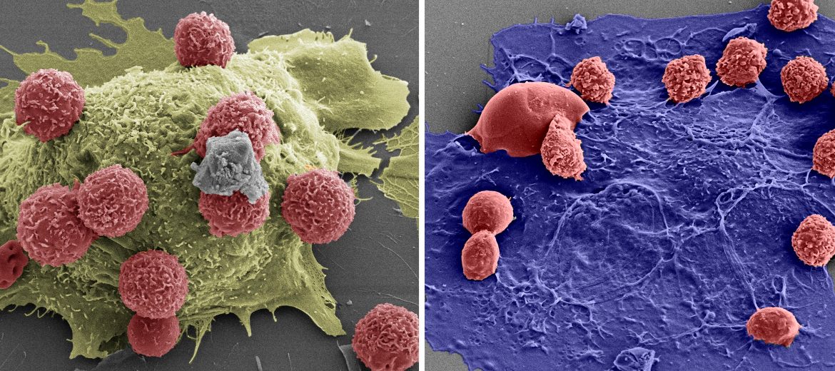
Prof. Fraser has used the imaging capability of the scanning electron microscope (SEM) in the AMMRF at USyd that shows a much lighter shade of grey for heavy elements, like gold, enabling clear visualisation of the gold-labelled proteins. His labeling experiments revealed that two distinct types of islands were present in the bone marrow. Each island has a central cell called a macrophage surrounded by the developing RBCs. Islands with a domed macrophage appear to be a niche for the earlier stages of differentiation whereas the flat islands appear to support later stages of RBC development and their eventual release into the blood.
This new understanding of RBC developmental processes will help interpret the errors that occur in RBC diseases.
J. Yeo et al., 2016, Microsc. Microanal. 22.
SEM images of the domed (left) and flat (right) EBIs. The developing red blood cells are about 5μm in diameter.
November 27, 2016