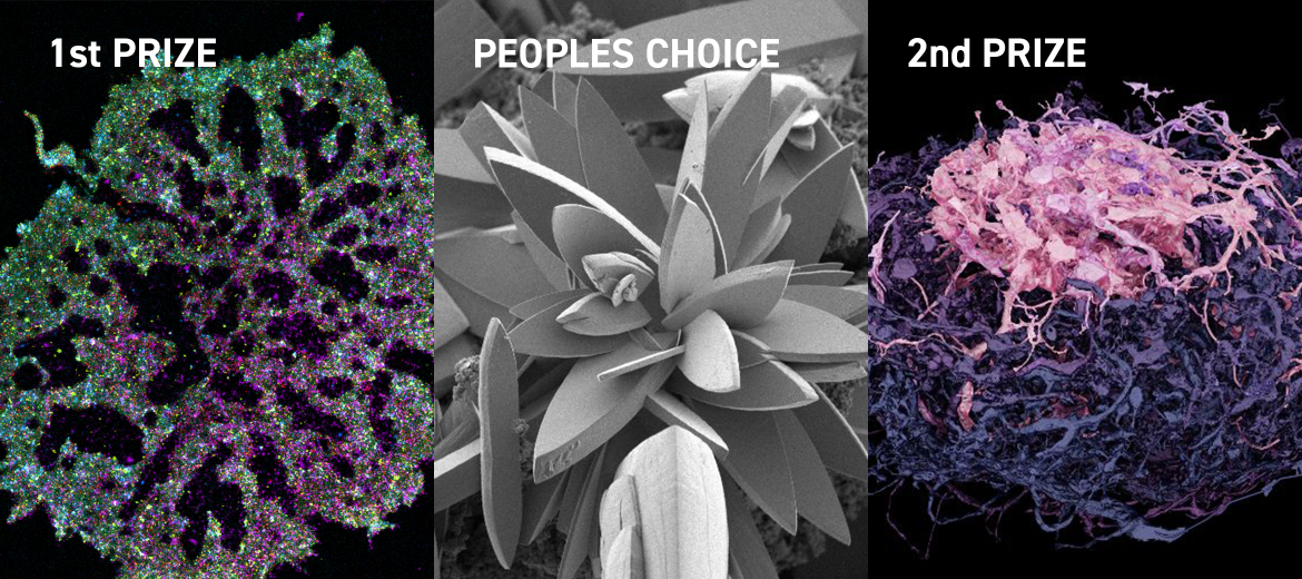
There were some great entries, mainly SEM and confocal and a smattering of other techniques. Thanks to all who entered and to the judges for their careful consideration of the microscopy presented. All images were anonymous at judging. Also, thanks to everyone who voted for the People’s Choice Award.
The overall winner was Dr Shirin Ansari, postdoctoral researcher at the EMBL Australia node for Single Molecule Science at UNSW Sydney, for her super-resolution image of an activated T cell captured using the single-molecule localisation microscopy technique, Protein PAINT. It shows five different components of the signalling cascade and offers a glimpse into the complex molecular organisation that underpins T-cell activation.
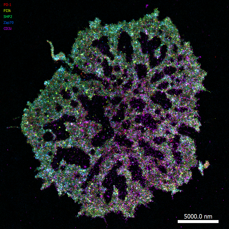
First prize winner – super-resolution image of an activated T cell by Dr Shirin Ansari
The second prize winners were Nicole Schieber, University of Queensland (UQ), and Valentin Gillet, Lund University, Sweden. Nicole is the Lab Manager of Electron Microscopy for Life-Science and Soft Matter at UQ’s Centre for Microscopy and Microanalysis. Their winning image was a three-dimensional serial blockface reconstruction of part of the neural circuitry in a Sweat Bee (Megalopta genalis). The reconstruction was generated with Blender from the individual segmented images covering a total volume of approximately 108x89x114 µm.
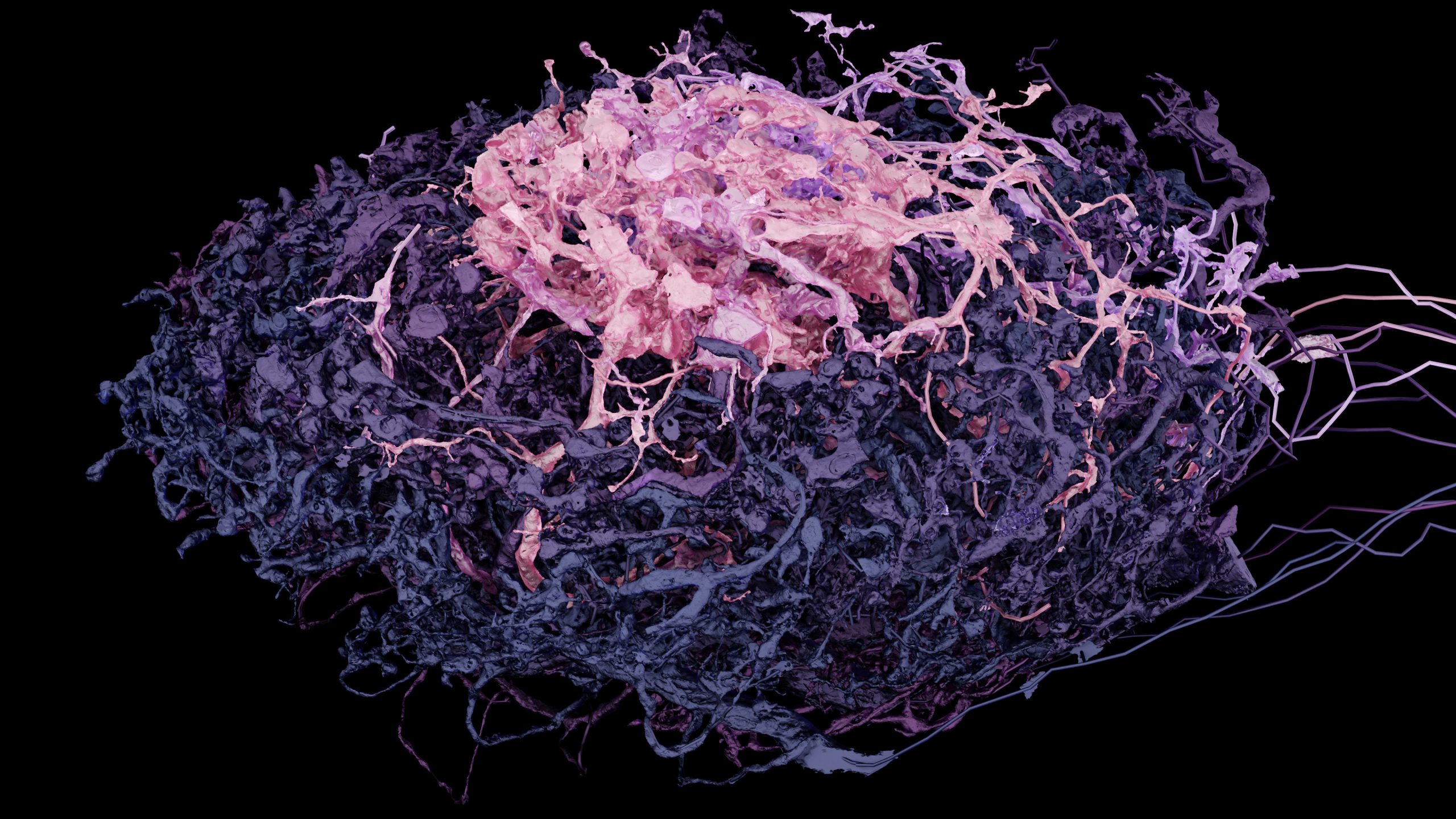
Second prize winner – three-dimensional serial blockface reconstruction of part of the neural circuitry in a Sweat Bee by Nicole Schiber and Valentin Gillet
The Peoples’ Choice votes gave a clear winner, with the prize going to Dr Heike Bostelmann, Platform Scientist at the Centre for Microscopy and Microanalysis at the University of Queensland for her beautiful SEM image of a rosette of copper sulphate crystals.
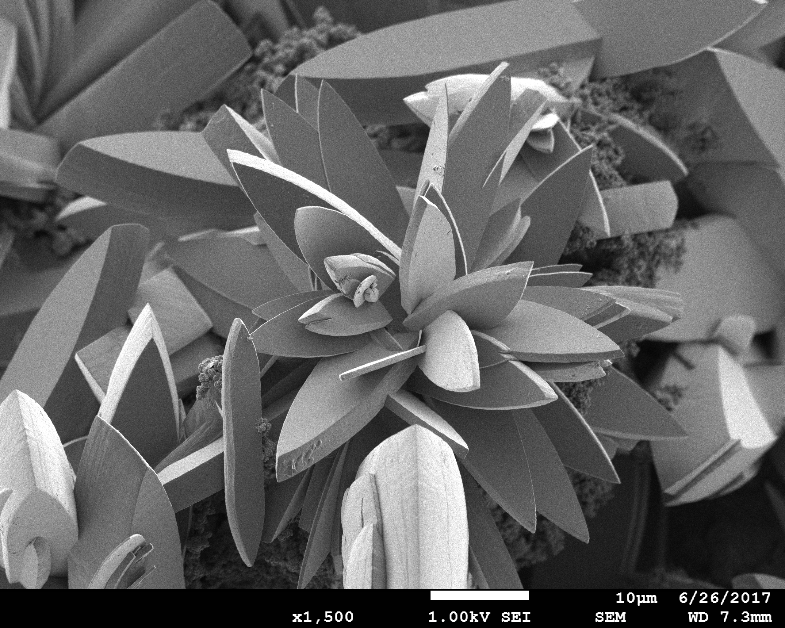
People’s Choice Winner – copper sulfate rosette by Dr Heike Bostelmann
February 14, 2025