For example, the scaffold can be wrapped around damaged nerves of people with acute trauma or neurodegenerative disorders. In many cases, nerve damage is permanent. This is changing, however, because this ‘internal bandage’ can provide support and growth factors to the tissue – allowing it to heal.
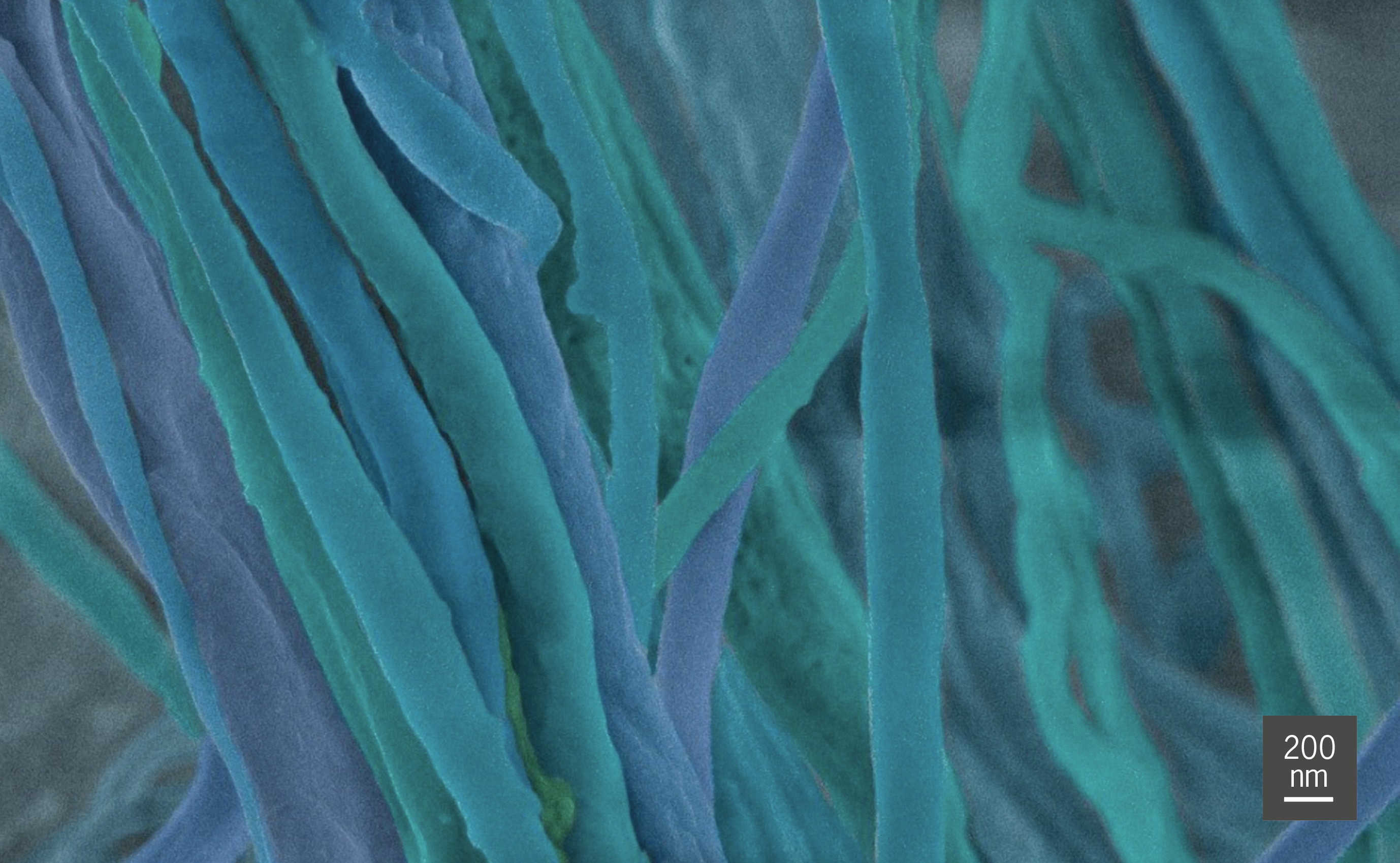
Ms Kiara Bruggeman at the Australian National University (ANU), under Dr David Nisbet’s supervision, produced a polylactic acid nanofibre scaffold using electrospinning. The fibre was then made functional by attaching proteins to it. To optimise the effectiveness of the scaffold, it is important to determine the fibre dimensions, orientation, consistency, surface characteristics and any undesirable artefacts. Ms Bruggeman used scanning electron microscopy (SEM) in the AMMRF (now Microscopy Australia) at ANU to do this.
The team used protein immobilisation techniques to trap various proteins on the scaffold: proteins such as interleukin 10, which guides the immune response to nerve injury, and brain-derived neurotrophic factor, which promotes neuronal differentiation and proliferation. This allows more healing and recovery because the trapped proteins remain active for longer than would proteins in solution, which degrade or diffuse rapidly.
Since neural injuries can lead to irreversible damage and disability, these findings could transform treatment – and therefore the future quality of life – for those with nervous system conditions.

Colour-enhanced SEM image of the nanofibre scaffold.
Colour-enhanced SEM image of the nanofibre scaffold.
October 24, 2014