Tooth enamel is a very tough material – the hardest in the body, and its nanoscale structure is extremely intricate. It is a biocomposite with 96 percent mineral phase, one percent enamel proteins and three percent water. This leads to a unique combination of strength and fatigue resistance. The mineral is hydroxyapatite (HAP) organized into orderly bundles of nanowires. It is known that magnesium ions regulate HAP crystallisation by stabilising its precursor, amorphous calcium phosphate (ACP). Knowledge of the atomic-scale distribution of Mg ions within the precursor ACP in mature human dental enamel would provide much needed information for a better understanding of enamel formation.
Dr Alex La Fontaine and Prof. Julie Cairney from the University of Sydney have used atom probe tomography to produce the first-ever three-dimensional positional maps of the atoms. These are the first direct observations of a Mg-rich ACP phase between the HAP nanowires in mature human dental enamel. The research team also observed Mg-rich elongated precipitates and pockets of organic material among the HAP nanowires.
Because the Mg-ACP phase at the enamel boundaries is susceptible to dissolving in acidic environments, the researchers propose that decay occurs via dissolution along the enamel rod boundaries. This knowledge will inform further research to develop strategies to enhance remineralisation, slow the progression of or prevent caries, or even restore lost dental enamel.
A. La Fontaine et al., 2016, Science Advances, 2 : e1601145
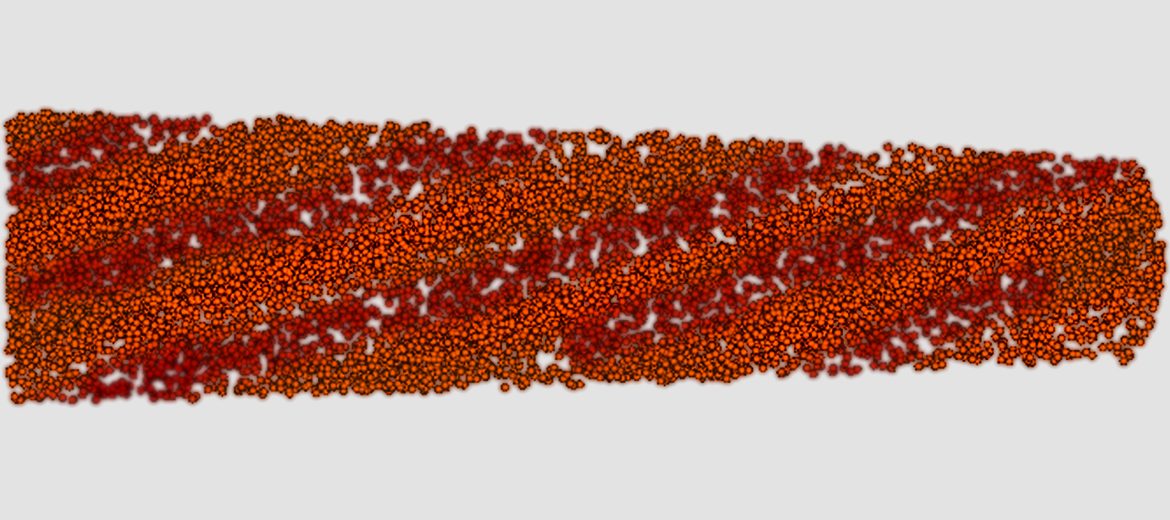
Atom probe data showing the regular arrangement of increased Mg concentration throughout the enamel. Length of sample approx: 350 nm
November 27, 2016