They are thought to provide stretch resilience to muscles and blood vessels and manage a turnover of lipids in the membrane. Reduction or lack of the caveolar proteins leads to problems ranging from tumours and neuronal diseases to life-threatening abnormalities in skeletal and heart muscles, lipodystrophy and metabolic imbalance. These clinical symptoms are linked to abnormalities in caveolae formation but the cause is not understood at the molecular level.
Dr Oleksiy Kovtun at the University of Queensland (UQ) is attempting to decipher the molecular architecture and cooperative action of proteins with lipid membranes in caveolae formation. He isolated a set of caveolar proteins called cavins. Biophysical analysis revealed that they formed a large hierarchical protein assembly. To understand the structure of this assembly Dr Kovtun used X-ray crystallographic techniques. This revealed the nature of the first product in the hierarchy, but the form of larger cavin assemblies remained elusive.
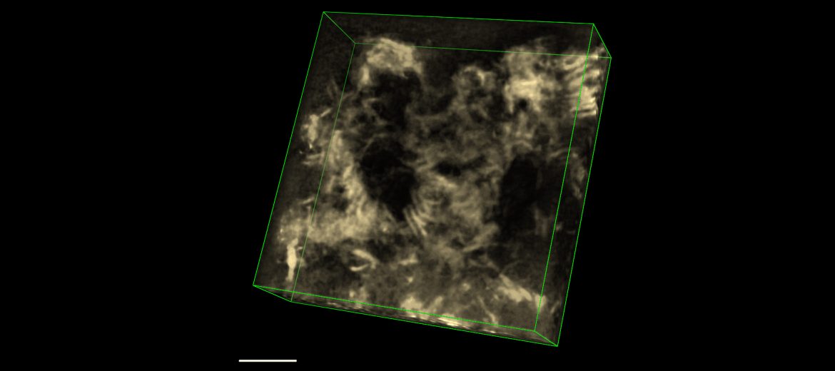
By using electron microscopy (EM) in the AMMRF (now Microscopy Australia) at UQ, he was able to make the breakthrough that revealed that the principal building block of cavin complexes are rod-shaped assemblies and that they have a specific arrangement in the final coat. EM imaging also suggested that cavin alone is sufficient for the characteristic membrane curvature of caveolae. These pioneering structural studies will help us to understand how caveola are formed and the causes of caveolae-linked pathologies.
2014. Kovtun, et al. Dev. Cell 31, 2014.
3D electron tomography image showing rod-shaped cavin assemblies. Scan the QR code to see a tilt series of cavin rods.
October 24, 2014