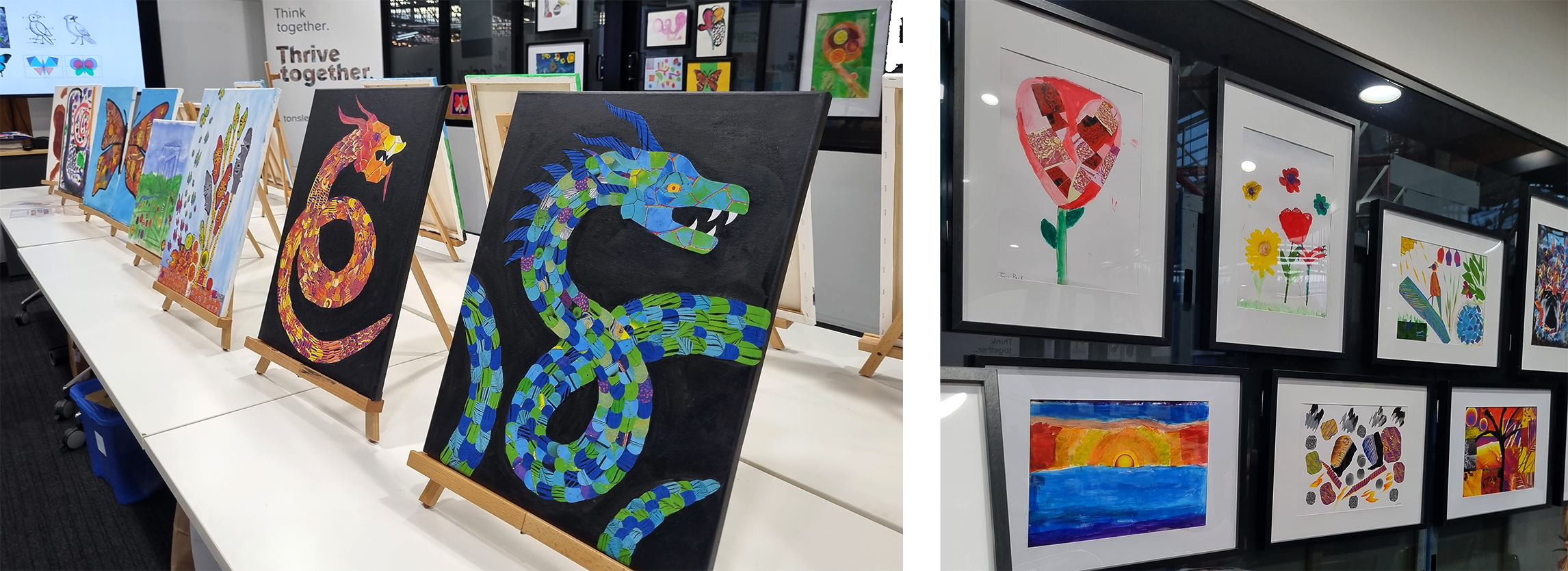
Some of the mixed-media artwork displayed in the Tonsley Pod.
The project, funded by a $7,000 science-art grant from Inspiring SA, involved hosting a series of community workshops to create nature-themed artworks for display at the ‘Seeing Things Differently – SmART Science’ exhibition on Feb 23rd for the Adelaide Fringe at Tonsley Innovation District.
Workshops used microscopy and X-ray images from Flinders Microscopy and Micro Analysis (FMMA)’s large volume micro-CT X-ray scanner and scanning electron microscope (SEM) and Micro-X’s portable X-ray instrument. You can read more about how Microscopy Australia’s South Australian facilities supported the development of Micro-X’s patented miniturised X-ray tubes here.
Participants were encouraged to create artworks combining the large-scale view of a nature object, such as a butterfly, with the small scale view obtained from microscopy. They could also make a nature-themed collage using any of the up to 700 coloured images available. Nature objects such as butterfly and dragonfly wings, feathers, leaves and seeds were used to tie in with the art of the nature artist, Erica Lee, who supported workshops.
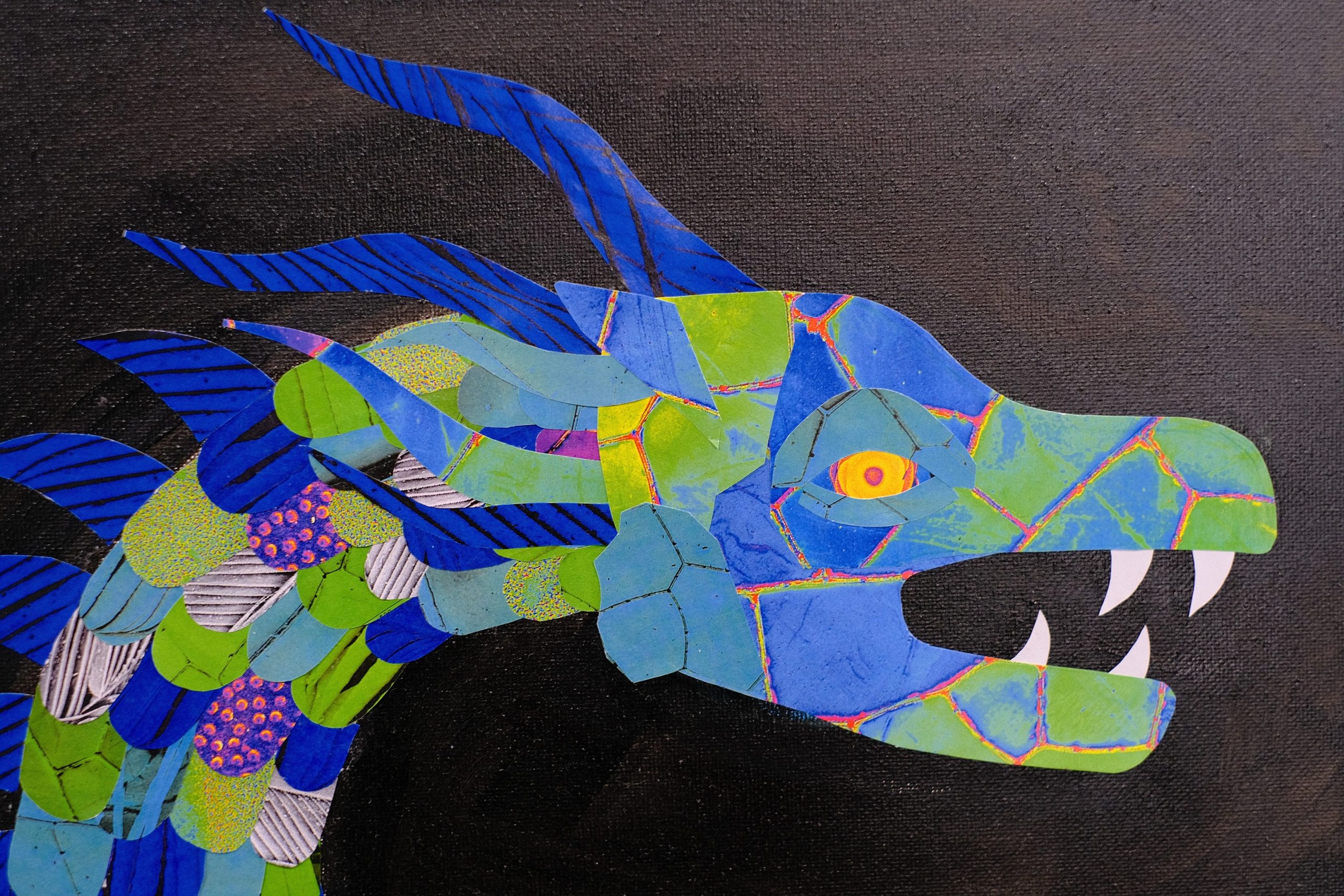
“Blue Dragon” by Olivia Gascooke uses SEM images of feather and gum leaf for the scales, a dragonfly wing for the face, an artichoke micro-CT X-ray for the eye and feather SEM for the horns and spines.
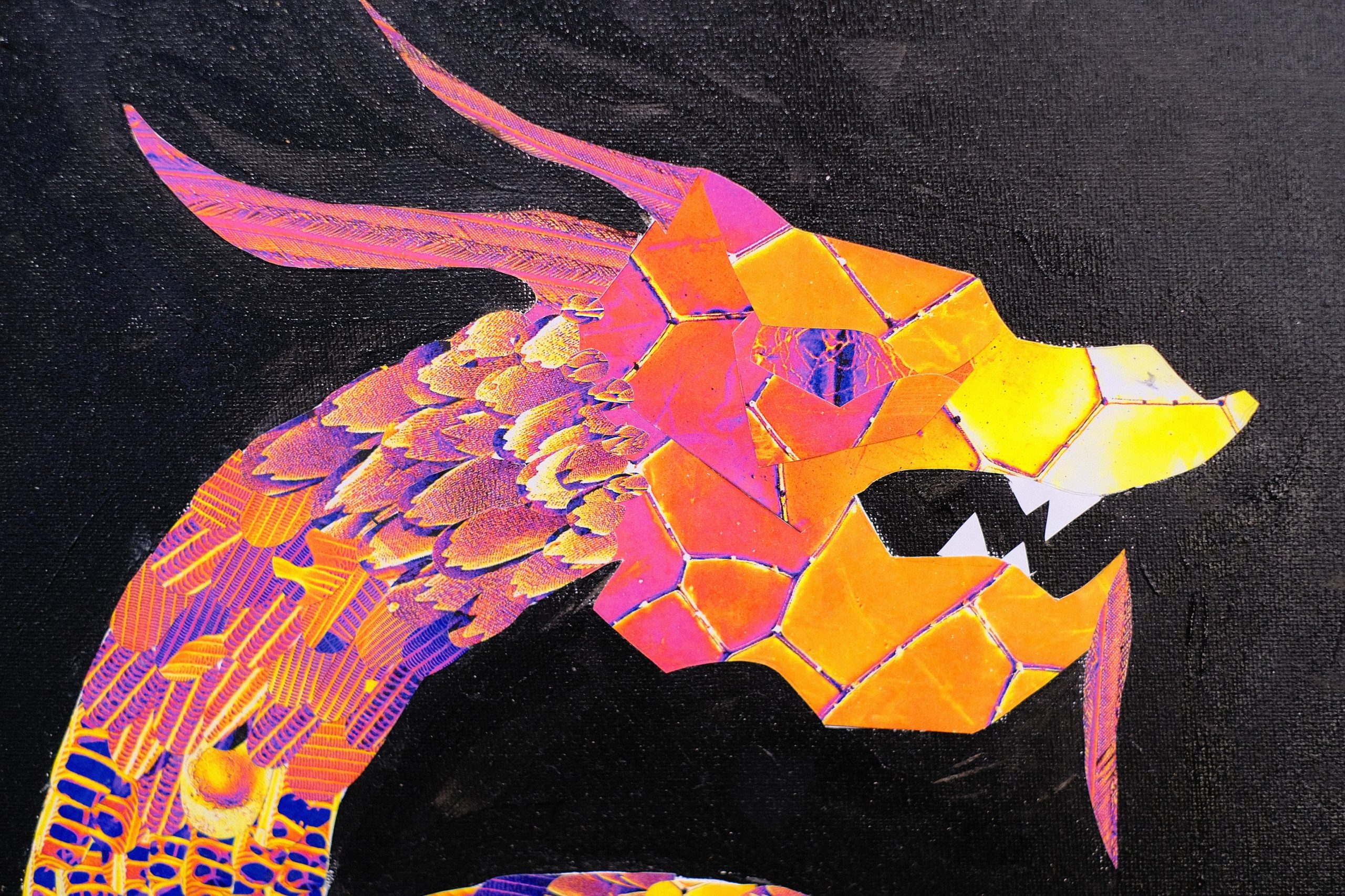
“Red Dragon” by Zali Gascooke uses coloured SEM images of a butterfly wing and gum leaf for the scales, dragonfly wing for the face, and feather for the horns and chin spike.
The result was an amazing variety of creativity. Over 110 artworks were created from 5 mixed-media and digital workshops with 60 participants ranging in age from 5 to 75 years. The exhibition included framed artworks and canvases, a digital display on the Flinders Tonsley screens and an augmented reality (AR) display trail through the Tonsley Garden.
Workshops included some of the science behind the microscopy images and why microscopy is useful for science and industry. For example, the micro-CT scanner can see inside objects non-destructively. The SEM shows high-level surface detail that helps with understanding why a dragonfly’s wing has anti-microbial behaviour, why water beads on a rose petal or how the structure of a butterfly’s wing plays with light. Science and industry can then try to mimic these structures to make bio-inspired photonic devices or self-cleaning surfaces that improve our lives.
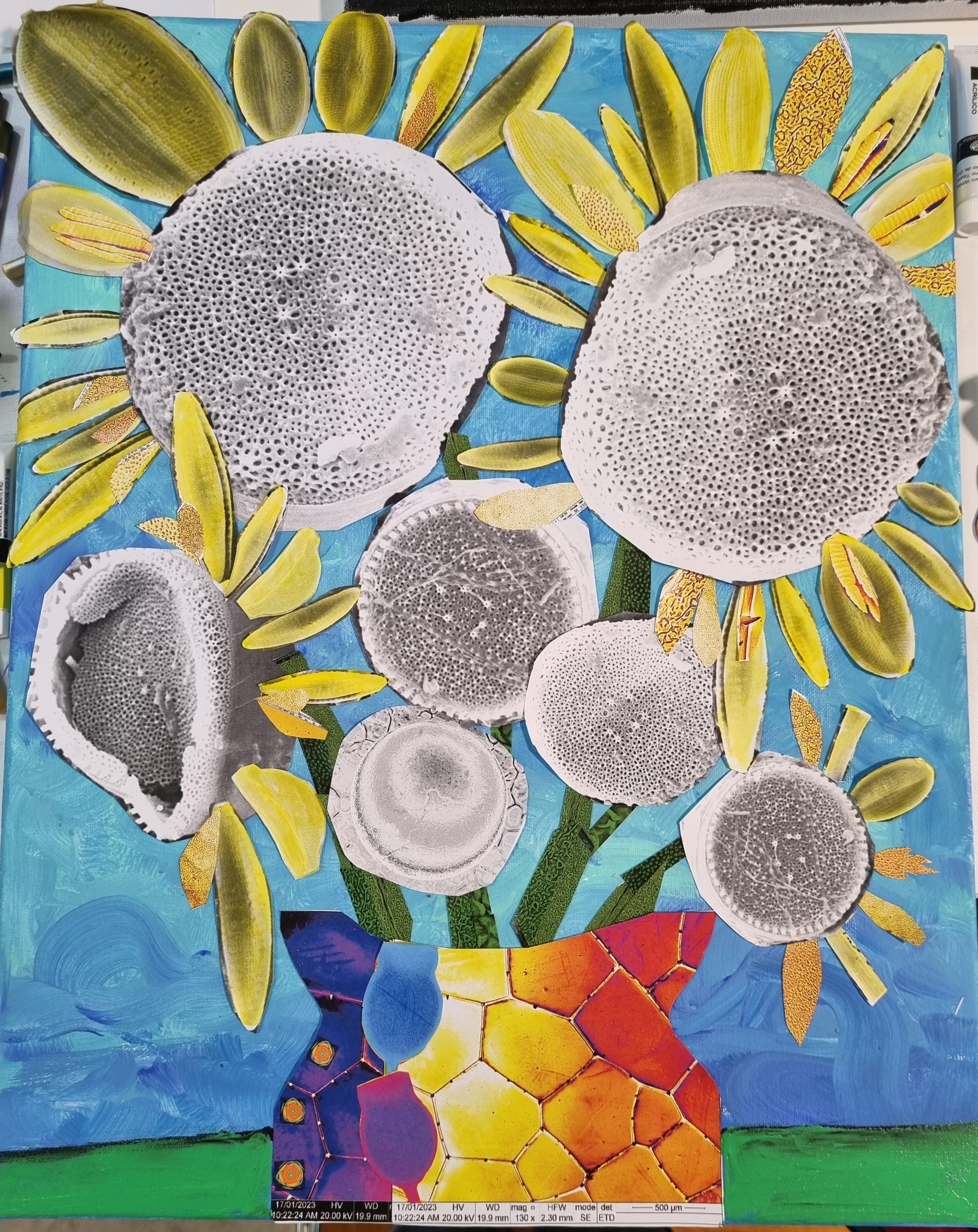
“Sunflowers” by Ula Alexander uses SEM of diatoms (courtesy of Prof Sophie Leterme) as sunflower centres and petals, also uses SEM of steel ball, gingko leaf, gumleaf with a dragonfly wing SEM vase decorated with gum nut and artichoke micro-CT images. SEM and Micro-CT images supplied by Flinders Microscopy and Microanalysis.
The “SmART Science” Project was led by Dr Ula Alexander, Microscopy Australia Industry Application Scientist at FMMA and Susanne Sahlos, Research Chemist from Micro-X. Thank you to Dr Sophie Rapagna and Dr Alex Sibley from FMMA for assistance with micro-CT and SEM image collection. Thank you to Evan Sahlos for supporting the augmented reality and digital workshop component of the project.
The AR trail and some of the artworks were also displayed at the Tonsley Science Fair on March 19th along with a hands-on art activity as part of Micro-X and Flinders displays. Remaining artworks and augmented reality trail QR codes are now on display outside the FMMA Office.
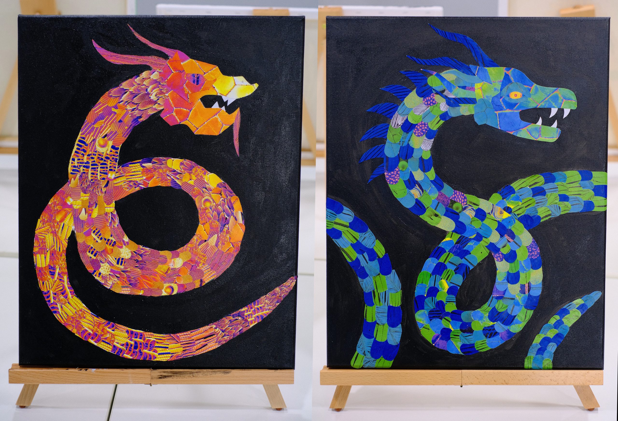
Full size of “Red Dragon” by Zali Gascooke (left) and “Blue Dragon” by Olivia Gascooke (right).
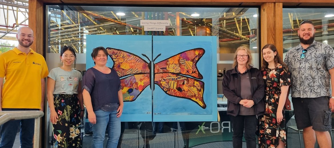
Exhibition helpers Evan Sahlos, Erica Li (Lee), Ula Alexander, Susanne Sahlos, Rebecca Santrac and Brodie Condon with a large version of “Butterfly” by Stella Hart and Lorraine Hart, that uses coloured scanning electron microscope (SEM) images of a butterfly wing, feathers, gum leaf and micro-CT X-ray images of a gumnut and artichoke.
March 30, 2023