Despite the potential of extracellular vesicles as selective biomarkers in medical diagnostics, little is known about their microstructure. Because the vesicles are typically 30-500 nanometres in diameter, transmission electron microscopy is often used to provide adequate resolution when imaging the vesicles. However, TEM sample preparation is tedious and time consuming and the highly sensitive vesicles can lose their structures upon contact with harsh chemicals,
at high temperatures or under vacuum.
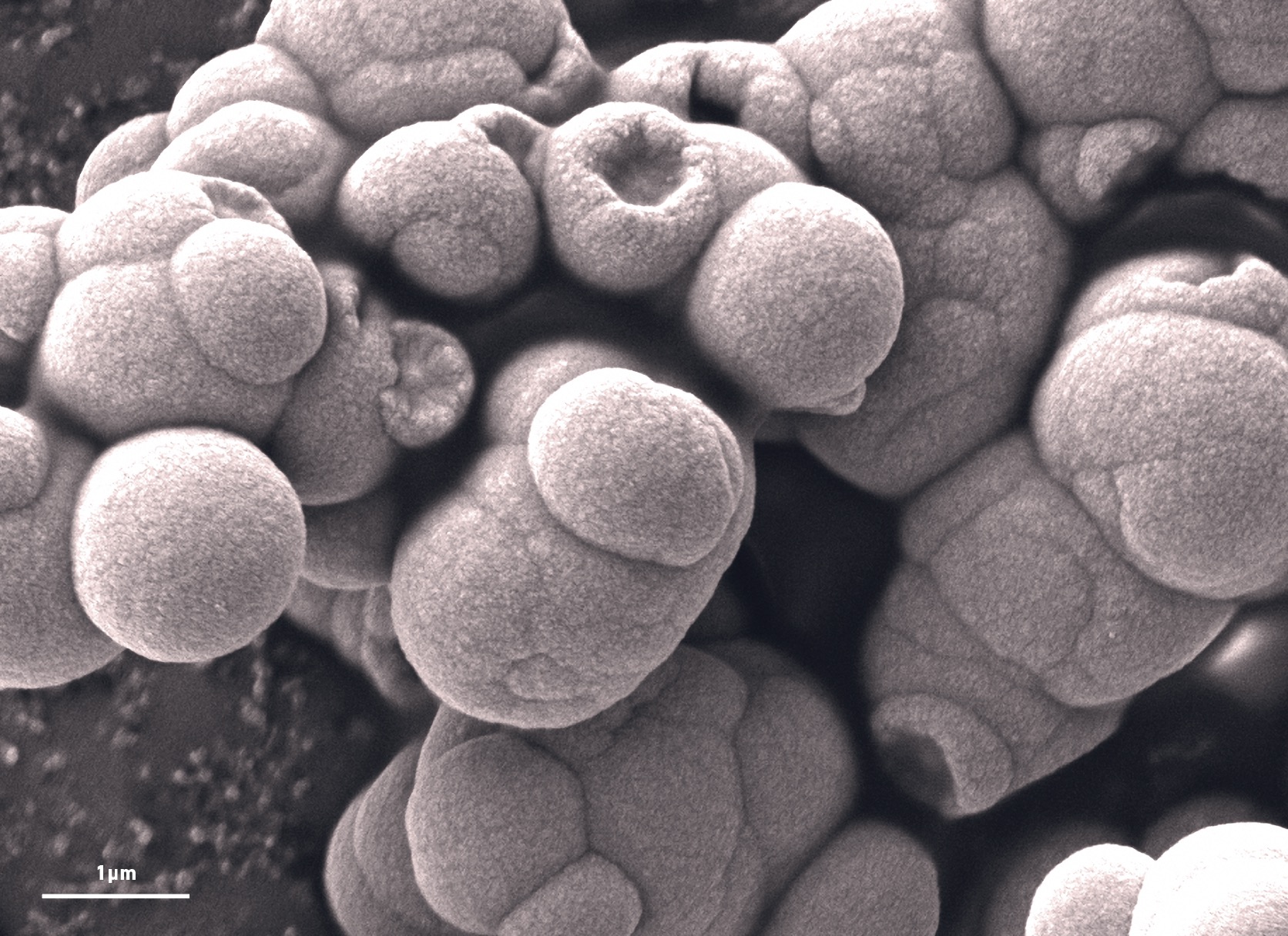
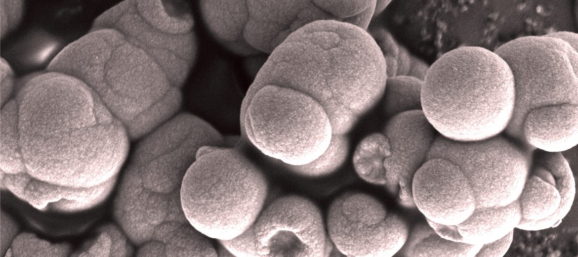
Scanning electron micrograph of extracellular vesicles after freeze-drying.
To develop a more efficient imaging method Dr Wojciech Chrzanowski and PhD student Sally Yunsun Kim at the University of Sydney (USyd), together with the Microscopy Australia biological specimen preparation specialist Naveena Gokoolparsadh, developed a unique, innovative method for preparing extracellular vesicles for ultra high resolution scanning electron microscopy (SEM). They used freeze-drying to immobilise the vesicles on polyethylenimine-coated ThermanoxTM glass slides. This gave high quality samples that provided excellent resolution in the SEM at Microscopy Australia’s USyd node.
This method proved to be highly successful, with the team obtaining high resolution structural images. Other aspects of this work are the subject of a current patent application.
July 26, 2017