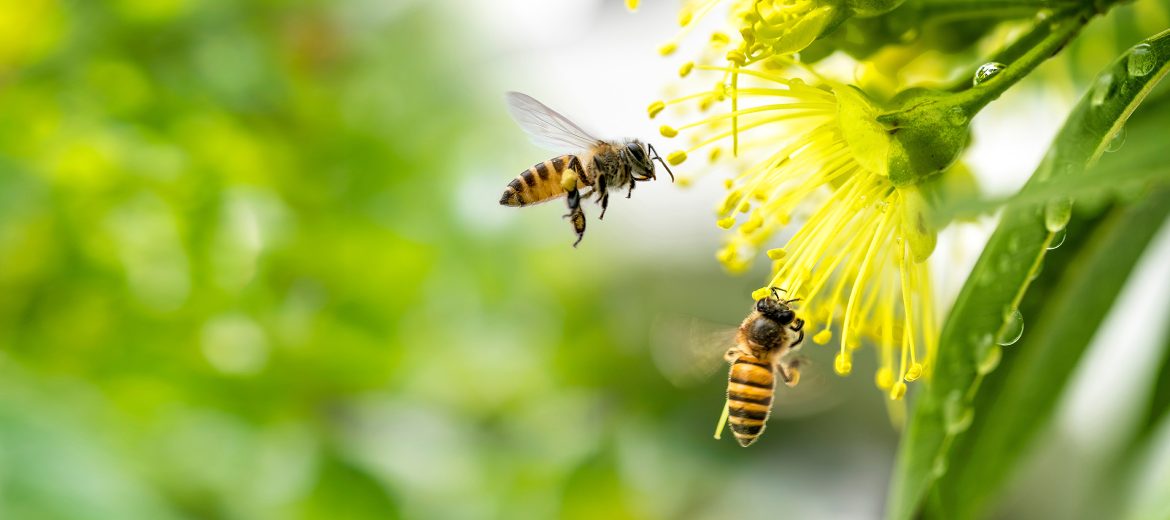
These observations inspired researchers at UNSW Canberra to learn more about how this complex system works with a view to designing securely attached, self-pumping medical devices for delivering hormones and drugs.
Dr Fiorella Ramirez-Esquivel and Dr Sridhar Ravi from the Australian National University (ANU) and UNSW Canberra have built on historical descriptions of the stinger by using the complementary microscopy techniques of scanning electron microscopy and micro-CT at our ANU facility. This was combined with high-speed video, producing data that allowed the complex 3D anatomy and motions of the stinger to be understood. Earlier work enabled by our University of Queensland facility, showed that the lancet component of the bee stinger is strengthened with manganese.
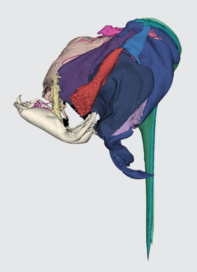
A 3D reconstruction of a stinger from micro-CT data showing the different components in different colours. The stinger is 2.5mm long.
The entire stinger is only 2.5mm long yet consists of a complex arrangement of moving parts including sections of the cuticle that pierce the highly elastic skin of vertebrates; muscles to insert the stinger and pump the venom; nerves to coordinate the process; and glands to make and store the venom and the alarm hormones that alert other worker bees.
The researchers are now developing 3D printed larger prototype needles and pumping devices to identify the key design features that give the stinger its unique properties. This will help to design simplified bioinspired devices that can then be manufactured in smaller sizes.
F. Ramirez-Esquivel & S. Ravi, iScience 2024
DOI: 10.1016/j.isci.2023.107103
January 4, 2025