Yellow fever is re-emerging as a serious disease threat, moving into urban areas of tropical and sub-tropical Africa and South America. It is caused by a mosquito-borne flavivirus that can lead to severe liver disease and, in some cases, death. Although researchers have been investigating the yellow fever virus for over a century, its structural details remained elusive. There are no approved drug treatments for yellow fever, and it is mainly controlled by mass vaccination campaigns with a non-infectious version of the virus. However, the strain currently used to make the vaccine is not proving to be as effective against emerging virulent strains of the disease as it has been in the past. A better understanding of the viral structure could help reveal the cause and a potential solution to this problem.
The first high-resolution structures of yellow fever virus have been revealed by cryo-electron microscopy (cryo-EM) at Microscopy Australia’s University of Queensland (UQ) facility. A collaborative team led by Drs Summa Bibby, James Jung, Naphak Modhiran and Prof. Daniel Watterson at UQ, created a hybrid flavivirus based on the harmless Binjari mosquito virus. The Binjari core was coated with surface proteins from different strains of yellow fever virus.
The cryo-EM data revealed surprisingly stark differences between the surface appearance of the vaccine strain and the virulent strain currently infecting people. The surface proteins of the two strains were known to differ by several amino acids and the cryo-EM structural studies made it clear that just one of these was responsible for the large structural changes in the two strains.
Cryo-EM is a powerful tool with which researchers could identify not only the structure of the yellow fever virus surface protein but also how the 180 individual copies of it fit together on the outside of each virus particle. Cryo-EM also enabled the team to determine how and why that one amino acid caused the altered structure.
In the vaccine strain, all the different surface proteins lock together in a stable arrangement, mediated by that one amino acid. However, in the virulent strain, that single amino-acid difference prevents the proteins from locking together properly. This leaves them unconstrained and free to wave around on the surface. This in turn presents a surface that looks quite different to what the patient’s immune system has been prepared for by the vaccine. It is therefore not surprising that the current vaccine is less effective against the virulent strains.
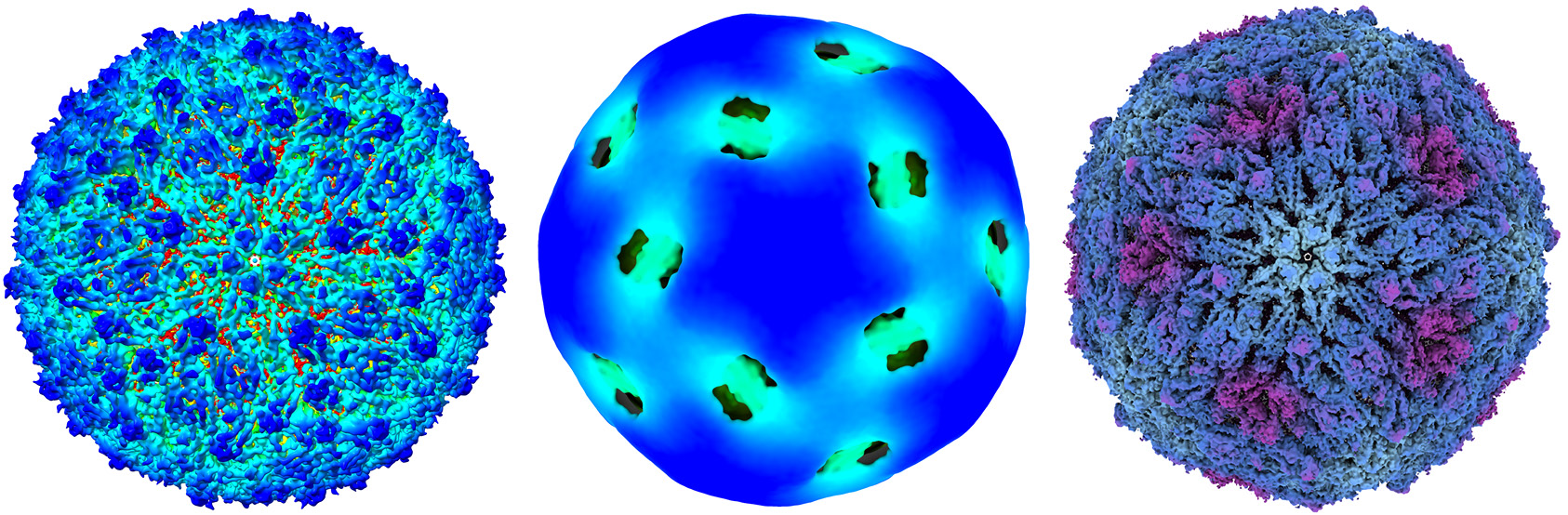
Left: Detailed cryo-EM structure of the vaccine strain. ~40 nanometres wide.
Centre: Cryo-EM of the virulent strain. The surface is too flexible to be imaged in detail by cryo-EM.
Right: Cryo-EM of the virulent strain with the mutant amino acid replaced by that from the vaccine strain.
These findings have provided crucial knowledge that will enable the research team to design more effective yellow fever vaccines and ones that don’t rely on a live, although inactive, virus. Potentially this knowledge could also enable development of valuable treatments. These outcomes will help to:
S. Bibby et al., Nature Communications 2025
DOI: 10.1038/s41467-025-63038-5
May 5, 2026