To get a better understanding of how this occurs at the atomic scale, materials engineers worked with dentists and bioengineers to map the exact composition and structure of tooth enamel at the atomic scale. The University of Sydney researchers, Prof. Julie Cairney and Dr Alex La Fontaine have pinpointed some of the nanoscale elements that govern the behaviour of our teeth.
Using atom probe tomography in Microscopy Australia at the University of Sydney, their work produced the first-ever three-dimensional maps of human tooth enamel showing the exact positions of atoms critical in the decay process.
The new knowledge of detailed atomic composition has been published today in the journal Science Advances and has the potential to aid oral health and caries prevention.
Prof. Julie Cairney, said, “Dental researchers have known that certain trace ions are important in the tough structure of tooth enamel but until now it had been impossible to map them in detail. The structure of human tooth enamel is extremely intricate and while we have known that magnesium, carbonate and fluoride ions influence enamel’s properties, scientists have never been able to capture enamel’s structure at a high enough resolution to see the ions’ relationships to each other in 3D.”
“What we have found are magnesium-rich regions between the hydroxyapatite nanorods that make up the enamel. This means that we have the first direct evidence of the existence of a proposed amorphous magnesium-rich calcium phosphate phase that plays an essential role in governing the behaviour of teeth.“
Dr Alex La Fontaine, said, “We were also able to see nanoscale ‘clumps’ of organic material, which indicates that proteins and peptides are heterogeneously distributed within the enamel rather than being present along all the nanorod interfaces, as was what was previously suggested. This mapping has the potential to inform new treatments designed around protecting against the dissolution of this specific amorphous phase and provide an understanding of how to help the remineralisation of tooth enamel.”
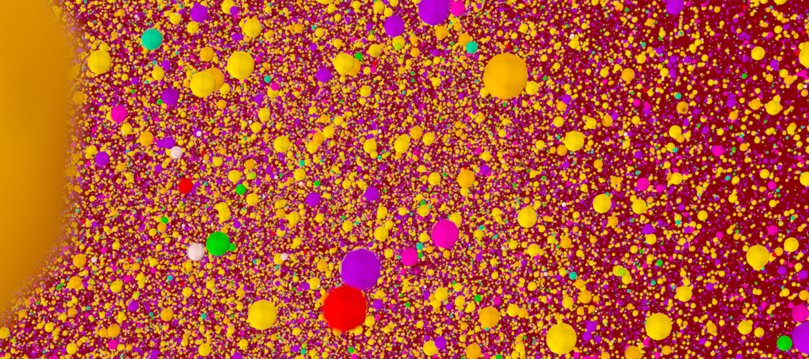
In the image, balls of different colours represent atoms of different elements as below.
 |
C – Black Ca – Orange Cl – Purple F – Turquoise H – White Mg – Green N – Tennis-ball colour Na – Yellow O – Red P – PinkAtomic radii of all elemental species in the image are accurate and to scale, relative to one another and to the size of the sample as a whole. |
Human dental enamel at atomic scale
September 8, 2016