The combination of different types of data acquired over different length scales and over time generates deeper understanding and more significant outcomes. This Profile contains many examples where multimodal microscopy enabled discovery and innovation.
The collaborative structure of the AMMRF (now Microscopy Australia) facilitates multimodal microscopy through instrument proximity and the critical mass of expertise available to researchers. This ecosystem is also immensely valuable in developing innovative new instruments, techniques and analysis tools.
Dual- or multi-modal instruments that automatically correlate data from the same region of the same sample are now becoming available. Around the AMMRF, staff work with microscope manufacturers to help develop these emerging technologies. Such integrated instruments are an exciting frontier in microscopy and promise increased efficiency in acquiring highly accurate correlated data.
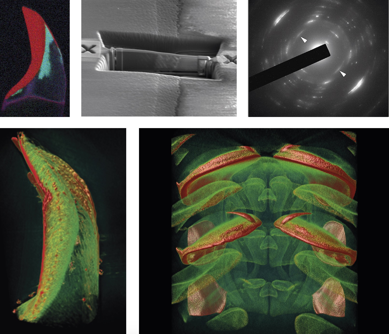
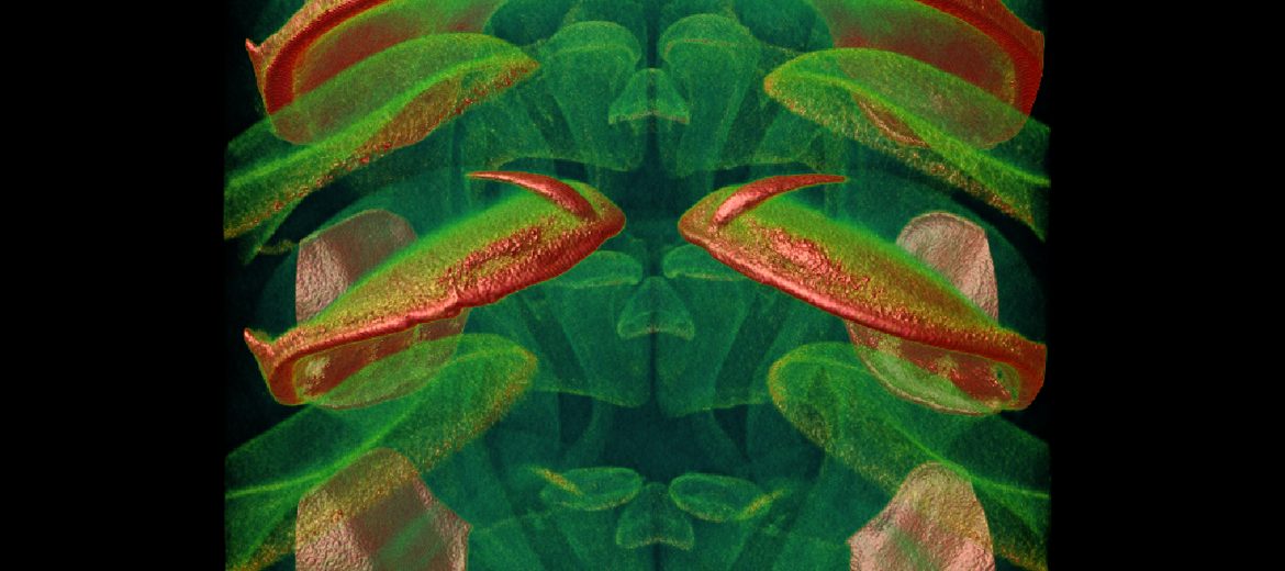
Left to right: 1. Elemental analysis in SEM showing formation of minerals. 2. Selected area cut out using FIB to be used for diffraction. 3. Diffraction in the TEM to identify mineral structure. 4. X-ray nanotomography for 3D nanostructure. 5. X-ray microtomography for 3D structure of anatomical arrangement.
Dr Jeremy Shaw is studying chiton teeth – a very strong natural composite material. He used transmission electron microscopy (TEM) for structural studies and energy-filtered elemental mapping at high magnification, along with scanning electron microscopy (SEM) and elemental analysis (EDS) at the tissue level. Focused ion beam (FIB) milling was used to extract small slices from selected areas of the tooth for diffraction studies. X-ray nano- and micro-tomography revealed the 3D structure of the teeth. Together these techniques give insights into the process that forms this tough biomineralised, iron-reinforced material at ambient temperatures.
November 27, 2016