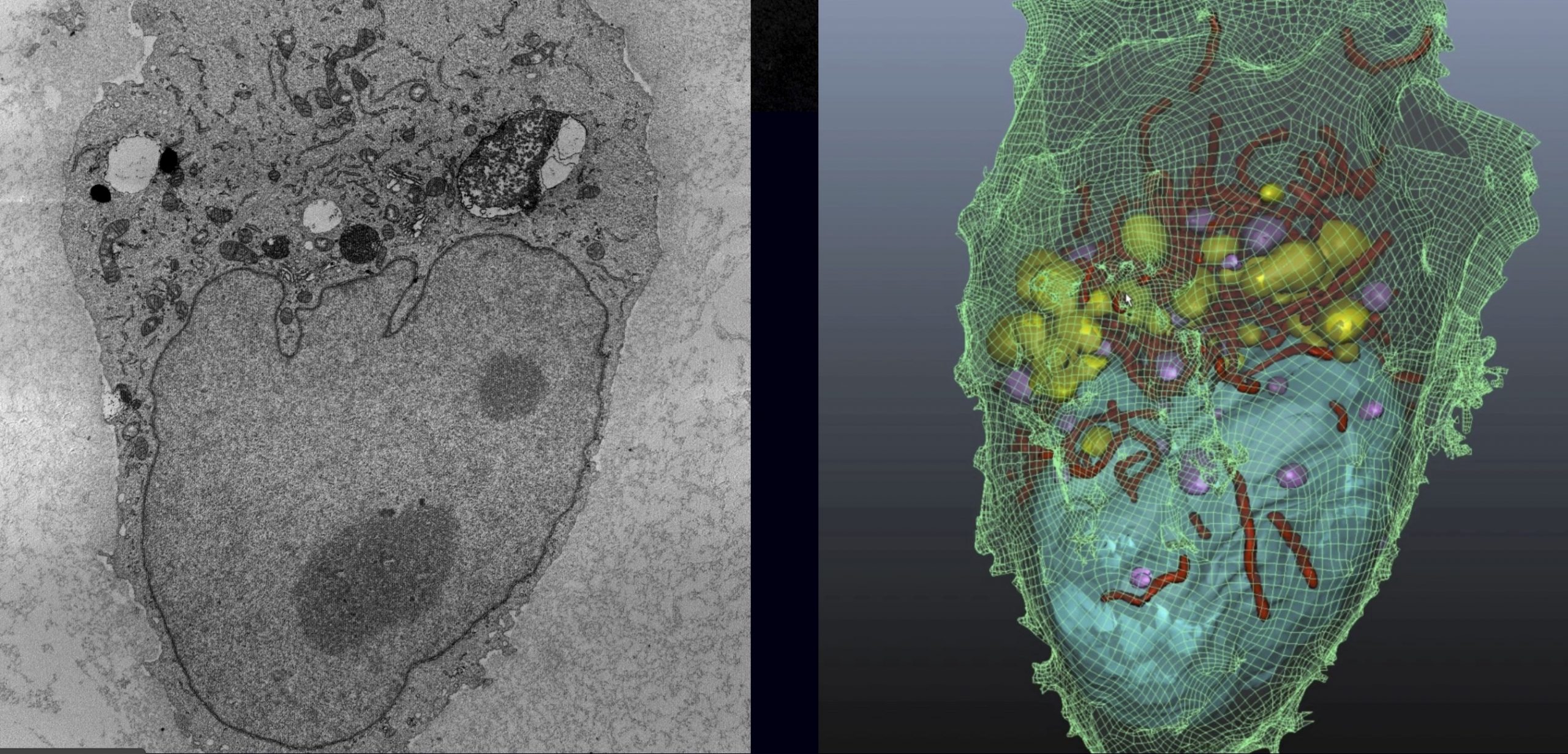
3D visualisations make it far easier to conceive the relative positions of components in the nanolandscapes of cells and molecules. To take real 3D microscope data and combine it with virtual reality (VR) technology could help researchers make new discoveries by seeing cellular structures and processes in a new and more tangible way. This in turn could lead to new insights and ‘eureka moments’.
Collaboration:
This Australian team developed Journey to the Centre of the Cell using high-resolution electron-microscope data from the AMMRF (now Microscopy Australia) at UQ to reconstruct a human breast cancer cell in digital 3D. The result is an immersive, interactive VR experience.

By wearing VR headsets scientists can navigate the ‘landscape’ of the cell – including the nucleus, mitochondria and endosomes. The visualisation aims to speed up the science discovery process by showing how nanoparticle drugs are absorbed by cancer cells. Researchers find it extremely useful to be able to walk around the cell, put their head inside a mitochondrion and have a look around. It also opens up the possibility of taking molecules that are mutated in different forms of cancer and putting them in the context of a virtual reality cell, so researchers can watch how they work.
Dr Johnson is focusing on how to deliver drugs inside tiny nanoparticles. He is using the VR cell to help improve delivery by finding ways to direct these nanoparticles to precise locations within the cell.
November 27, 2016