Research has shown that bone marrow lesions (BMLs) directly beneath the knee cartilage (subchondral bone) correlate with symptoms and disease progression but can only seen using magnetic resonance imaging (MRI). University of Adelaide (UAdel) PhD student Ms Dzenita Muratovic and her supervisors, Dr Julia Kuliwaba and Prof. David Findlay, wanted to characterise BMLs at the tissue level and if BMLs seen in different MRI imaging modes are characterised by different tissue morphology.
Ms Murotovic used MRI to localise BMLs then characterised the subchondral bone microarchitecture in fifty knee specimens from patients undergoing total knee replacement for osteoarthritis. Assisted by AMMRF (now Microscopy Australia) staff, Ms Muratovic used the AMMRF at UAdel to develop a specific X-ray microtomography imaging and analysis protocol for large, complex specimens. They were the first to use this protocol to analyse the 3D microarchitecture of BMLs. Their images showed different degrees of change in the subchondral bone. One BML type represents an earlier stage of osteoarthritic disease progression, whereas another is likely to represent a later stage and is, therefore, harder to treat.
The team proposes MRI of BMLs as a specific biomarker to identify individuals at risk of progressive osteoarthritis, and as a way to monitor therapy. Given the prevalence of osteoarthritis, new ways of monitoring the condition will be clinically important.
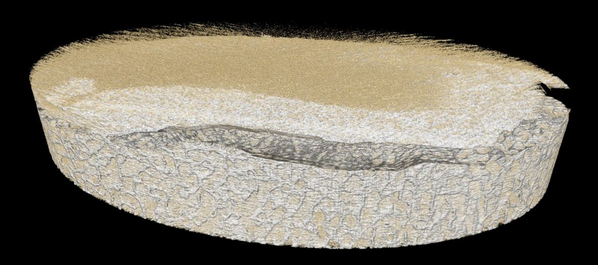
3D reconstruction of X-ray tomography data of a whole tibial plateau showing loss of cartilage.
October 24, 2014