Efficient drug delivery methods are necessary in the management of respiratory diseases. A key property of successful treatments is the ability of carrier particles to efficiently deliver the drugs deep into the lungs.
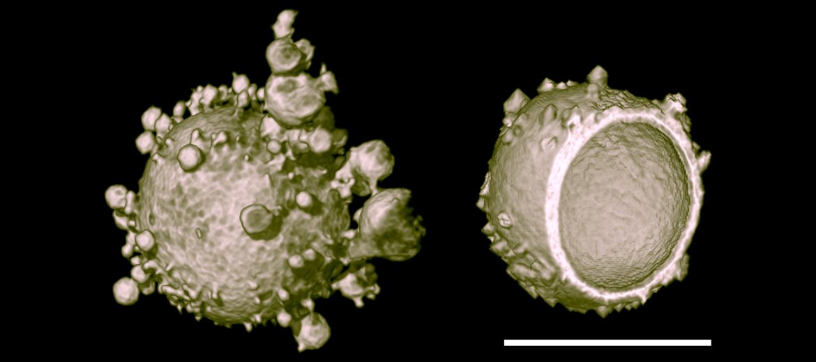
Using X-ray nano computed tomography (nano-CT) in the AMMRF (now Microscopy Australia) at the University of Sydney, researchers led by Prof. Kim Chan have been able to visualise the internal structures of porous pharmaceutical particles in a non-destructive manner at resolutions approaching 50 nanometres. Previous studies have required sectioning of the particles through microtomy or focused ion beam microscopy. These methods have left researchers unsure as to whether the delicate internal features had been impacted by the pre-imaging treatment.
Prof. Kim’s team has imaged a variety of different drug carrier particles, including sugars, polymers, proteins and lipids. The technique allows visualisation and quantification of the internal porosities with confidence that the data obtained is representative of the true nature of the particles.
Overall, X-ray nanotomography enables characterisation of drug delivery particles in greater detail. The team plans to continue using the quantitative capabilities of the X-ray nanotomography system to explain and predict the aerodynamic behaviour of their particles. This will assist them to develop more efficient drug delivery products to improve the quality of life for patients suffering from respiratory diseases.
X-ray nanotomography reconstruction of a PLGA particle. Scale bar is 18 μm. Scan the QR code to see a 30-micrometre diameter multi-porous particle.
October 24, 2014