Finding effective treatments for genetic disease is a long but essential process. A range of muscle diseases, including some muscular dystrophies, are caused by mutations in genes controlling the formation of caveolae. Caveolae are nanoscale pits on the surface of muscle cells.
Evidence accumulating from Prof. Rob Parton’s research at the University of Queensland (UQ), suggests that these caveolae could act as a reservoir of extra membrane to ease tension when the cell surface is stretched. This hypothesis was put to the test.
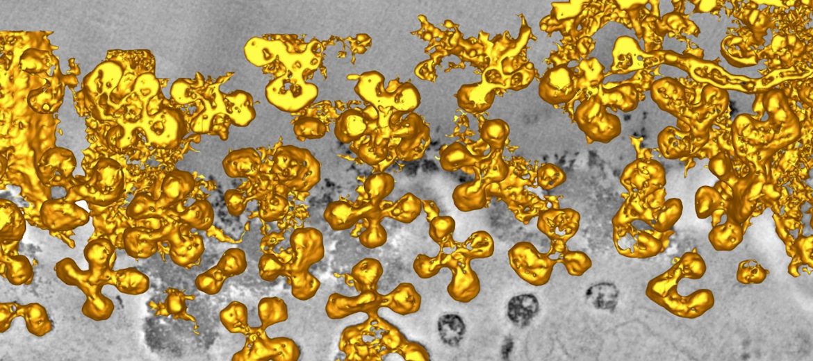
Dr Harriet Lo, working with Prof. Parton, compared muscle cells from normal mice to those from mice genetically modified to prevent caveloae formation through the deletion of the cavin-1 gene. By using 3D electron microscopy in the AMMRF (now Microscopy Australia) at UQ she found that caveolae occupied around 50% of the normal muscle cell surface and were predominantly assembled into multi-lobed rosettes. Increased membrane tension caused these rosettes to disassemble. Muscle fibres lacking caveolae showed a loss of cell membrane organisation, abnormal internal tubules, and increased sensitivity to membrane tension. This was all overcome when the cavin-1 gene was added back into the muscle cells of the mutant mice.
Following this, the team imaged living zebrafish embryos. In these embryos, disruption of caveolae led to cell membrane damage but only after vigorous muscle activity. It also seems likely that caveolae play a part in muscle repair.
Taken together their results demonstrate that caveolae, and the genes that produce them, are key to an inbuilt muscle protection system.
H. Lo et al., 2015, Journal of Cell Biology 210 (5).
This foundational knowledge is essential for building our understanding of normal muscle function and muscle diseases.
Surface-rendered reconstructions of caveolar rosettes in normal muscle fibres. Each caveola is approximately 50 nanometres in diameter.
November 27, 2016