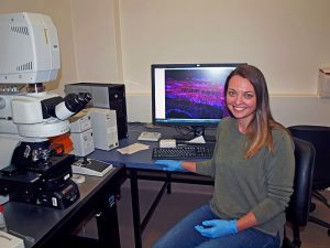
PhD student Kara Levin, working at Adelaide University’s Waite Research Institute, used their Microscopy Australia facilities to combine novel specimen preparation, confocal microscopy and high-resolution 3D imaging to reveal how cereal cyst nematodes hijack water and nutrients in wheat roots.

Kara Levin at the microscope
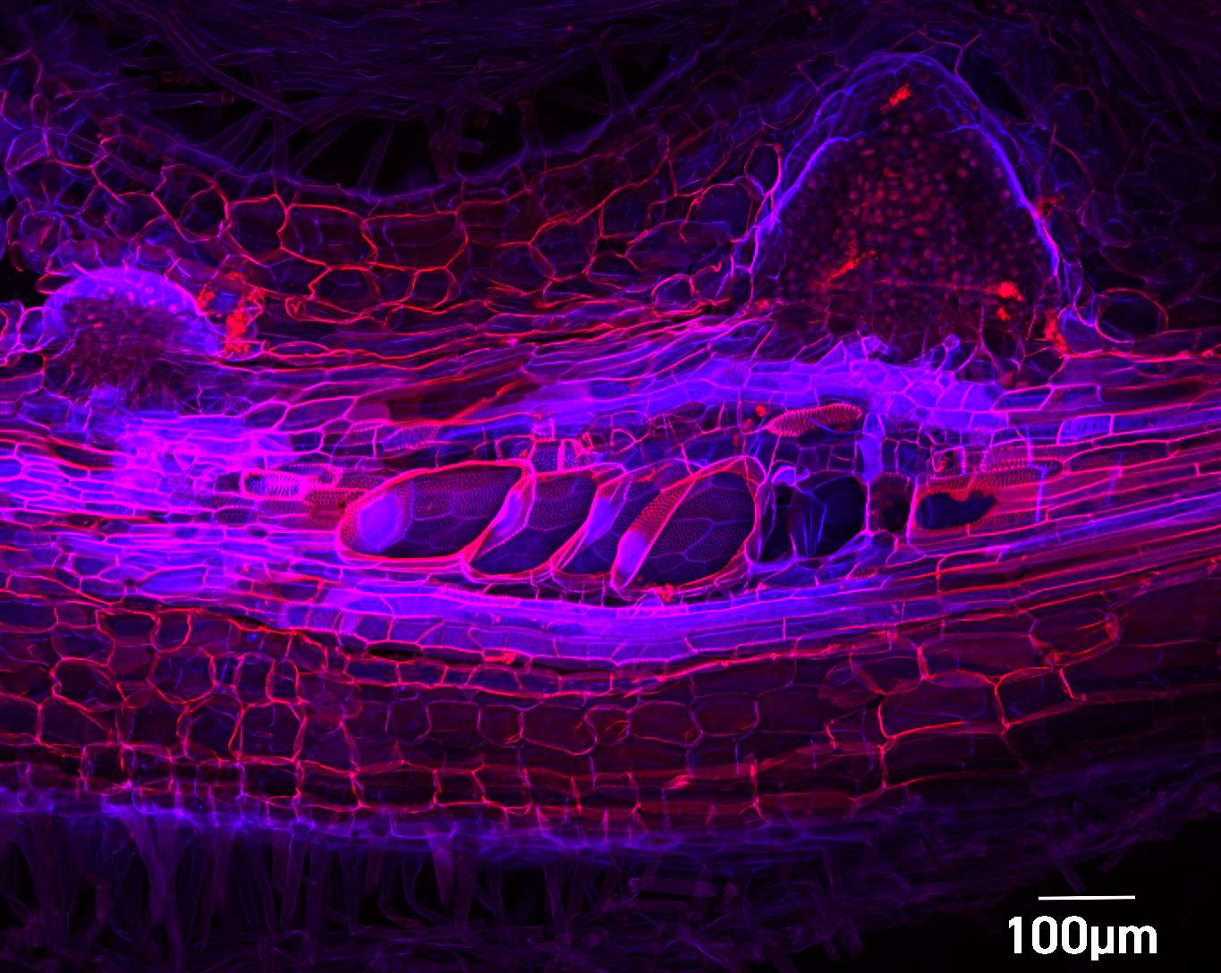
Comprehensive microscopic investigation of nematode feeding sites is complicated by their variable size, shape and depth within root tissue. The techniques used by Ms Levin in her research, and published in the journal Scientific Reports, allowed her to look deep within CCN-infected wheat roots and generate 3D models of nematodes and their effects on plant tissues. Rotational videos provided clear views of the unique anatomy of feeding sites, with sufficient resolution to reveal cellular-level spatial relationships among nematodes, feeding sites and the host vascular tissues that transport water and mineral nutrients. This approach revealed striking and previously unreported remodelling of developing metaxylem vessels after infection by CCN. By hijacking the classical developmental pathway leading to programmed cell death of some cells, xylem vessels can be kept alive for use by the parasite.
Ms Levin says that although microscopy has been used to investigate this system for decades, this 3D approach is the first to show that xylem cells in infected roots become short and fat ‘bubbles’ rather than long and narrow ‘tubes’. This is likely to impede water transport within the roots. Her supervisor, Prof. Diane Mather says this information is “important in determining resistance mechanisms in wheat that could be exploited in breeding programs”.

Confocal image of infected wheat root
3D model of nematode feeding site
June 30, 2020