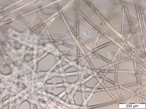
 Optical microscopy (right) and staff expertise at the University of South Australia are being used to develop methodologies, bespoke equipment and to test the quality and effectiveness of locally manufactured personal protective equipment.
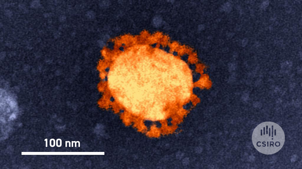
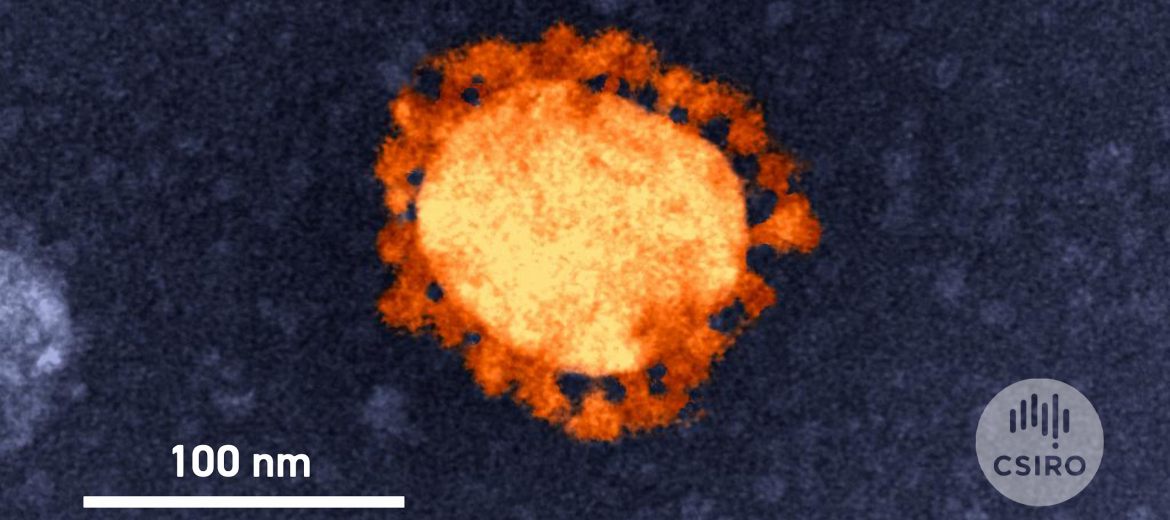
Colour-enhanced transmission electron microscope (TEM) image of the SARS-Cov-2 virus, which causes COVID-19. It was taken on the TEM in the high-containment lab at the CSIRO’s Australian Centre for Disease Preparedness (ACDP), a Microscopy Australia linked lab.
Colour-enhanced transmission electron microscope (TEM) image of the SARS-Cov-2 virus, which causes COVID-19. Taken on the TEM in the high-containment lab at the CSIRO's Australian Centre for Disease Preparedness (ACDP), a Microscopy Australia linked lab.
July 8, 2020