Dr. Haibo Jiang from the University of Western Australia (UWA) with collaborators from the National Physical Laboratory, UK and GSK developed a method to conduct high-resolution correlative backscattered electron and nanoSIMS imaging that shows structural and chemical information on drug distributions on the same section of the same cell. This method allowed visualisation of the internalisation of the drug amiodarone by lung macrophages and showed that it accumulated in internal compartments called lysosomes.
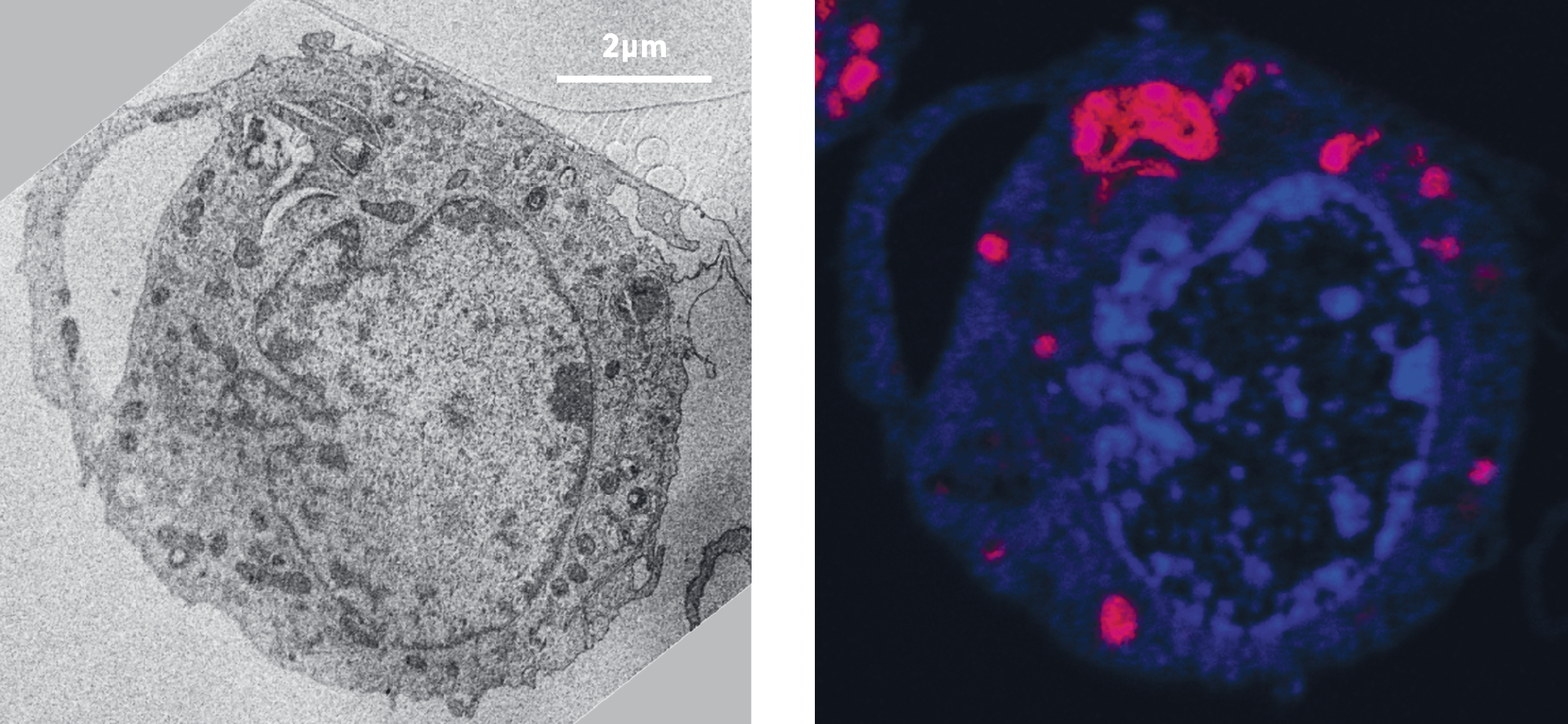
Correlative back scattered electron (left) and nanoSIMS imaging (right), showing the morphology of the cells, and distribution of amiodarone (red) in lysosomes.
Amiodarone is a drug used to treat and prevent irregular heartbeats. This family of drugs is able to easily cross cell membranes, making them very attractive from a pharmacological point of view. However, most of these drugs, including amiodarone, have been reported to induce phospholipidosis, a disorder leading to inflammation and dysfunction of a variety of cells and tissues. Using the method developed at UWA, the researchers also showed, with high-magnification correlative images, the first visual evidence of amiodarone-induced phospholipidosis and the colocalisation of amiodarone and membrane structures in the lysosomal lamellar bodies.
July 25, 2017