Skin to nerve cells
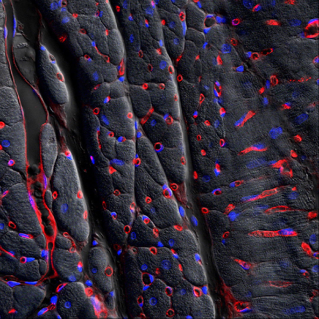
Red areas are cells that line capillaries feeding the heart muscle (the dark background area). The blue dots are cell nuclei.
Visualised using confocal microscopy (to see the colours) superimposed with differential interference contrast microscopy (to see the tissue structure) by Dr Paul Monaghan, Dr Tracey Hinton, Di Green and Dr Kim Wark, CSIRO.
Size: this piece of tissue is 260 micrometres wide
This is part of a project by CSIRO scientists to look at innovative ways to deliver new kinds of RNA-based treatments to tissues that may have been infected with viruses or have particular genetic problems.